A team of researchers with the Center for Ultrasound Research & Translation (CURT), in the Department of Radiology at Massachusetts General Hospital, have reported a pair of studies exploring the use of machine learning to improve ultrasound imaging in liver disease. In a recent conversation, team member Abder-Rahman Ali, PhD, lead author of both of the studies, walked us through the findings of each.
Liver Segmentation in Ultrasound Images Using Self-Supervised Learning with Physics-inspired Augmentation and Global-Local Refinement
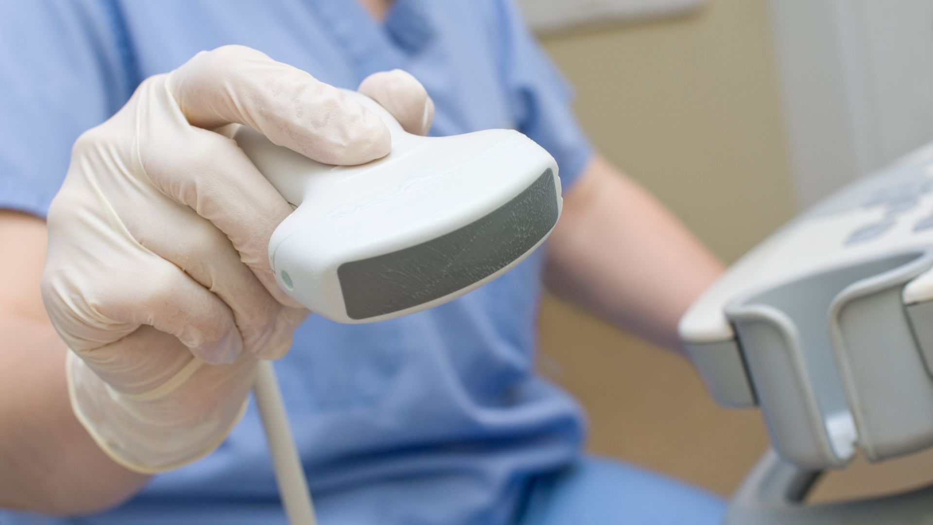
The growing prevalence of non-alcoholic fatty liver disease (NAFLD) has created a more urgent need for noninvasive means of diagnosis. Medical ultrasound can help, not least because it is widely available, portable and relatively inexpensive. For this application, though, the technique can be hampered by its low resolution — particularly with respect to liver segmentation, which is imperative for accurate diagnosis of NAFLD.
In a recent study, the CURT team sought to overcome this obstacle. The method they developed uses an integrated approach combining self-supervised machine learning, ENet fine-tuning, and physics-inspired data augmentations for improved liver segmentation in ultrasound images.
Ali outlines the benefits of the new approach. “The method employs the SimCLR model to learn feature representations from unannotated ultrasound images, reducing the need for large, annotated datasets, which are then fine-tuned using the ENet model,” he says. “Further, the incorporation of physics-inspired augmentations, specifically designed for ultrasound imaging, allows for a more accurate representation of real-world scenarios.”
The researchers reported the results of the study in June at the 36th annual Canadian Conference on Artificial Intelligence, describing the successful demonstration of the approach for liver segmentation in ultrasound images. The method yielded better segmentation performance than the comparative U-Net method, for example, and captured the complexities of the liver’s anatomy accurately.
The team is now looking to improve the quality of data used for training the downstream task (i.e., liver segmentation), Ali says, by utilizing a data-centric AI approach aiming to achieve better segmentation performance with less labeled data that has been systemically selected. They also plan to explore the use of a vision transformer (ViT) as the backbone of SimCLR, potentially enhancing the model’s learning capability and thus its performance in liver segmentation tasks.
Self-Supervised Learning for Accurate Liver View Classification in Ultrasound Images with Minimal Labeled Data
In a separate study, the CURT researchers tackled another, related challenge. For example, the AI models they employ are trained using abdominal ultrasound images, but not all such images contain liver views, so it is first necessary to detect those that do. Manual identification of liver views can be laborious and expensive, however, especially given the vast quantities of data involved.
Ultrasound imaging of the liver would therefore benefit from automated identification of ultrasound images with liver views prior to the training of AI models. To this end, Ali and colleagues developed the “SimCLR+LR” approach which combines contrastive self-supervised learning (SimCLR) with logistic regression (LR) to classify liver views in ultrasound images.
The researchers described the work in June at the 2023 IEEE / Computer Vision Foundation (CVF) Conference on Computer Vision and Pattern Recognition. “The two-stage method achieved high classification accuracy even with a limited number of labeled images,” Ali says. “It significantly outperformed state-of-the-art models, ResNet-18 and MLP-Mixer, and even reached similar performance to MLP-Mixer with only one labeled image per class.” He added that the findings emphasize the potential of self-supervised learning techniques, such as SimCLR, in situations where labeled data is hard to come by or otherwise expensive to obtain, “offering a promising substitute for traditional supervised learning methods.”
Looking forward, the researchers hope to expand the binary classification to a multiclass organ view identification. “We also aim to delve into Active Learning in order to make the model more efficient in handling unlabeled instances,” Ali says. “Furthermore, we intend to optimize the model’s performance and eventually deploy it in real-world clinical settings.”
The other authors of the two studies were Peng Guo, PhD, and Anthony E. Samir, MD, MPH, director of the Center for Ultrasound Research & Translation.